


Project members

Project
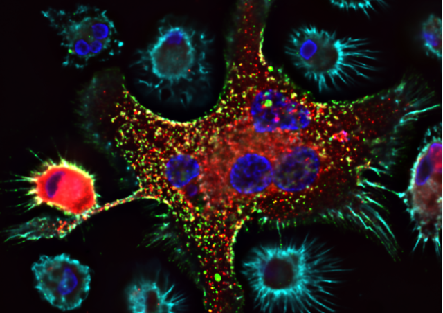
T cells can polarize and migrate in response to chemokines. These steps allow their entry into the lymph nodes but also promote their interaction with antigen presenting cells and thus their activation. For many years, we have been interested in identifying the signaling events triggered in T cells after antigen recognition or stimulation by chemokines. These questions are addressed with cell biology and real-time imaging approaches. Using biosensors, we can follow signaling events (calcium, cAMP, protein kinase A, ZAP70 tyrosine kinase activity...) at the single (or even sub)-cellular level and with good temporal resolution and correlate these events with cytoskeletal remodeling and the distribution of proteins of interest. We are currently focusing on the signaling events triggered in T lymphocytes in response to chemokines leading to the deformation (polarization) and migration of T lymphocytes.
Early signaling triggered in T lymphocytes after chemokine stimulation
Using dynamic imaging approaches and biosensors, we recently demonstrated that during chemokine-induced T cell migration, transient increases in cAMP were able to modulate the directionality of these cells by controlling the distribution of the actin cytoskeleton (Simao et al, 2021). This phenomenon, mediated by protein kinase A, promotes the exploratory behavior of T cells. We have also shown that protein kinase A is also important for the establishment of T cell polarization in response to chemokines. We are now trying to understand the signaling pathways involved. Is the polarity axis set up randomly? What are the cellular elements responsible for the symmetry breaking? Is it the protein kinase A itself or one of its substrates?
Funding